William K. Ovalle
Professor Emeritus
Director of Histology (1977-2010)
Muscular Dystrophy Association of Canada Postdoctoral Fellow (University of Alberta, Edmonton)
Ph.D. (Temple University School of Medicine, Philadelphia)
B.Sc. (St. Joseph’s University, Philadelphia)
Email: william.ovalle@ubc.ca
Wikipage: Essential Histology
Born in Panama, educated in the United States, William Keith Ovalle joined UBC’s Anatomy Department in 1972. Rapidly ascending the ranks to Professor in 1984, he is currently Professor Emeritus in the Faculty of Medicine. Besides teaching all branches of human anatomy and supervising graduate students and postdoctoral fellows, he was Director of Medical/Dental Histology for 33 years. With specialized training in both transmission and scanning electron microscopy, research endeavors span structural and histochemical aspects of normal and diseased skeletal muscle, muscle spindles, and muscular dystrophy. After initiating the department’s first graduate training program, he was appointed Head of Anatomy, and has served on various boards including the Canadian Association of Anatomists, Canadian Federation of Biological Societies, American Association of Anatomists, and Muscular Dystrophy Association of Canada.
His interest in anatomy – with special focus in microscopy – was influenced by several memorable role models: At age 10, his father gave him his first light microscope. At Temple University School of Medicine: thesis advisor (Dr. Steven Phillips) showed him how to fine-tune his first electron microscope – fostering a life-long fascination for cell/tissue ultrastructure. Neuroanatomy professor (Dr. Raymond Truex) adeptly displayed intricacies of the human brain. Noted embryologist (Dr. Robert Troyer) vividly pinpointed stages of early human development. Expert gross anatomists (Drs. Lorenzo Rodriguez-Peralta and Carson Schneck) highlighted clinical relevance of human dissection. During postdoctoral years at University of Alberta’s Department of Surgery, Dr. Richard Smith taught him how to micro-dissect muscle spindles in vivo and record intracellularly from intrafusal muscle fibers. In Anatomy at UBC: Professors Sydney and Constance Friedman created an exceptional academic atmosphere where traditional teaching and cutting-edge research were held equally and in highest regard; Dr. Chuck Slonecker sparked his interaction with student-learners in the classroom and laboratory; Drs. William Webber and Martin Hollenberg (former Deans of Medicine and notable microscopists) further nurtured his academic career every step of the way.
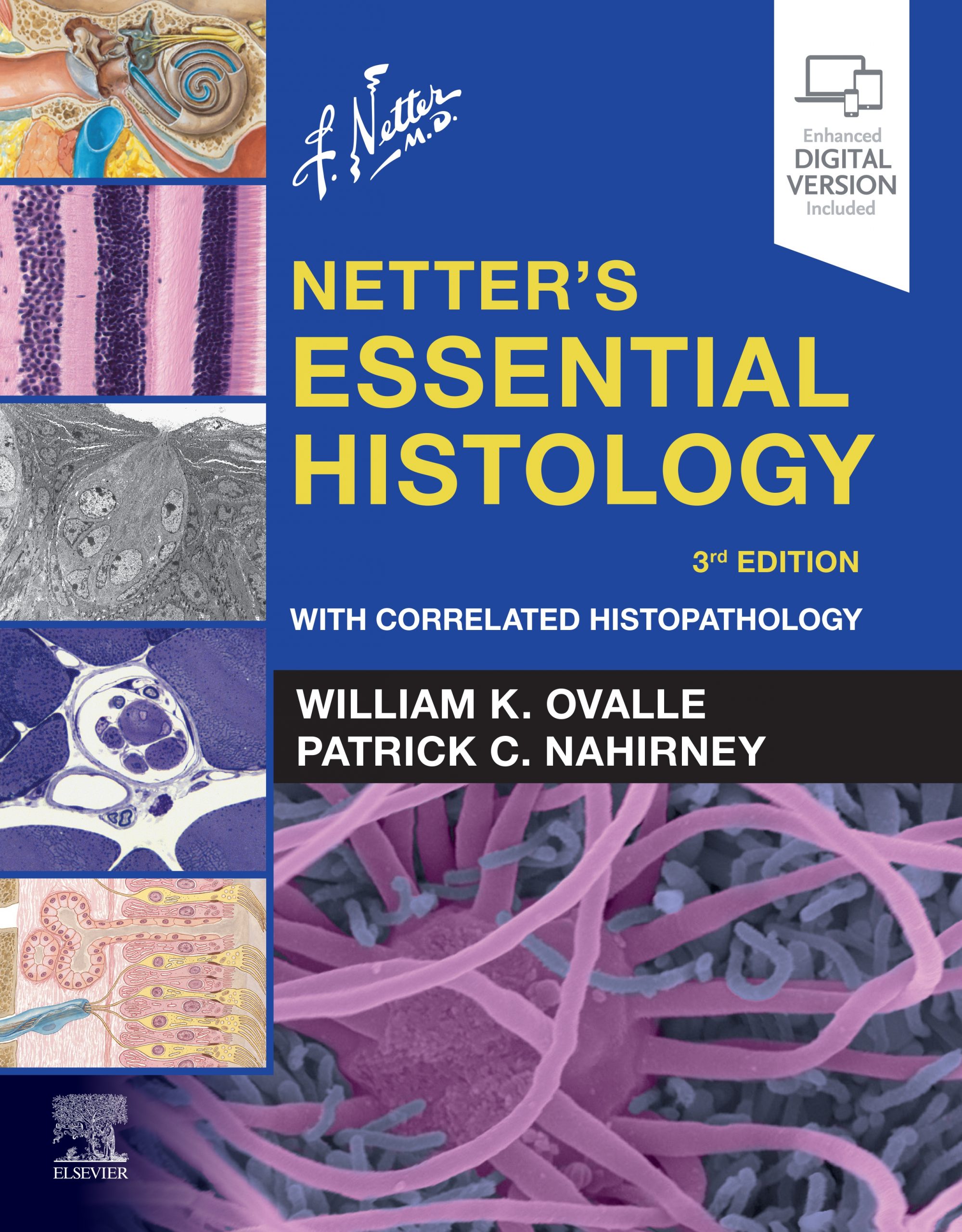
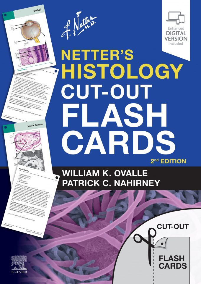
He is senior author of the popular textbook/atlas Netter’s Essential Histology published by Elsevier (with co-author Dr. Patrick C. Nahirney of the University of Victoria). Now in its 3rd edition (© 2021), previous versions were launched in 2008 (1st) and 2013 (2nd). Several foreign translated books (Portuguese, Turkish, Korean, Greek, Italian, Spanish) and Netter’s Histology: An Instant Review – First South Asia Edition (© 2014) have also been released. A new translation of the 3rd edition in Spanish (©2021) is now available. Book contents can be accessed online with an enhanced digital eBook version available on a variety of devices. New to each chapter in the 3rd edition are novel pages and salient images that focus on correlated histopathology. Author-narrated video overviews of all 20 chapters accompany a virtual slide library (digitized light microscopic slides and ’zoomifiable’ electron micrographs) and numerous ‘clinical points’ that highlight relevant information on disease and cellular dysfunction. Companion to the book is an elegant set of Histology Flash Cards used world-wide by students in medicine and the allied health professions; the newest updated version (©2020) consists of easy-to-use ‘cut-out’ cards.
As an educator with a long and rich history in microscopic anatomy, Ovalle has responded to the changing needs of his discipline – moving from a microscope focus – to pioneering the development of a virtual histology website for use in the expanded and distributed medical curriculum in British Columbia – the focus of other curricula around the world. This innovative and interactive website contains a Virtual Slidebox (> 500 original slides) and an EM Magnifier© (> 200 electron micrographs); these digitized, variable magnification images can be easily manipulated on the computer screen. With co-author (Dr. Nahirney), he continues to develop web-based instructional tools and multimedia learning programs in histology with clinical focus.
He has been recognized with the Killam University Teaching Prize (UBC’s highest educational award), several Medical Undergraduate Society Awards for Teaching Excellence, the Faculty of Medicine 50th Anniversary Gold Medal, the Panamerican Association of Anatomists Certificate of Merit, the British Medical Association Best Illustrated Book Award (2008), and Medical Media Review’s list of “25 Best Medical Books of All Time”. An Honorary UBC Medical Alumnus, he belongs to UBC’s Quarter Century and Tempus Fugit Clubs, and is especially honoured that three of his former graduate students became full-time faculty members at other medical schools in North America, and have won notable teaching awards at their institutions.
His leisure time is most things outdoors, spending it with his life-partner, Robert, and their adventurous Miniature Australian Shepherd, Opie. Besides ocean swimming and beachcombing, he also enjoys gardening and photography in upcountry Maui, crewing with the Maui (outrigger) Canoe Club, avidly attending yoga classes near the beach and participating in a monthly book club. He supports the Maui AIDS Foundation, Maui Humane Society, BC Insight Meditation Society, Hawaii Animal Rescue Foundation, and BC InspireHealth. He can be spotted in Vancouver’s annual Pride Parade, joining UBC Faculty of Medicine’s entry to this event. When not engaged in histology studies and writing, he is improving his golf and tennis scores with his extended family of friends.
Ovalle, W.K. Fine structure of rat intrafusal muscle fibers. The polar region. Journal of Cell Biology 51:83‑103 (1971)
Ovalle, W.K. and R.S. Smith. Histochemical identification of three kinds of intrafusal fibres in the cat and monkey based on the myosin ATPase reaction. Canadian Journal of Physiology and Pharmacology 50:195‑202 (1972)
Khosla, S., S.J. Tredwell, B. Day, S.L. Shinn and W.K. Ovalle. An ultrastructural study of multifidus muscle in progressive idiopathic scoliosis. Changes resulting from a sarcolemmal defect at the myotendinous junction. Journal of the Neurological Sciences 46:13‑31 (1980)
Ovalle, W.K. The human muscle tendon junction. A morphological study during normal growth and at maturity. Anatomy & Embryology 176:281‑294 (1987)
Anderson, J.E., B.H. Bressler and W.K. Ovalle. Functional regeneration in the hindlimb skeletal muscle of the mdx mouse. Journal of Muscle Research and Cell Motility 9:499‑515 (1988)
Patten, R.M. and W.K. Ovalle. Muscle spindle ultrastructure revealed by conventional and high resolution scanning electron microscopy. Anatomical Record 230:183‑198 (1991)
Nahirney, P.C. and W.K. Ovalle. Distribution of dystrophin and neurofilament protein in muscle spindles of normal and mdx-dystrophic mice: an immunocytochemical study. Anatomical Record 235:501-510 (1993)
Nahirney, P.C., P.R. Dow and W.K. Ovalle. Quantitative morphology of mast cells in skeletal muscle of normal and genetically dystrophic mice. Anatomical Record 247:341-349 (1997)
Goodmurphy, C.W. and W.K. Ovalle. Morphological study of two human facial muscles: orbicularis oculi and corrugator supercilii. Clinical Anatomy 12: 1-11 (1999)
Ovalle, W.K., P.R. Dow and P.C. Nahirney. Structure, distribution and innervation of muscle spindles in avian fast and slow skeletal muscle. Journal of Anatomy 194:381-394 (1999)
Books and Cut-Out Flash Cards:
Flashcards
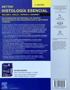
Ovalle, W.K. y P.C. Nahirney. Netter. Flashcards de Histologia. Spanish translation: Barcelona, Spain: Elsevier, 2021 https://www.elsevier.com/books/netter-flashcards-de-histologia/ovalle/978-84-9113-956-0
Foreign Book Translations
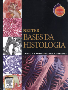
Ovalle, W.K. and P.C. Nahirney. Netter Bases Da Histologia. Portuguese translation: Elsevier/Saunders, Philadelphia 2008 (ISBN: 978-85-352-2803-8), 493 pages.
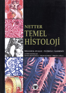
Ovalle, W.K. and P.C. Nahirney. Netter Temel Histoloji. Turkish translation: Elsevier/Saunders, Philadelphia 2009 (ISBN: 978-975-277-220-5), 486 pages.
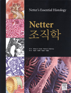
Ovalle, W.K. and P.C. Nahirney. NETTER 조직학. Korean translation: Elsevier/Saunders, Philadelphia 2009 (ISBN: 978-89-92589-59-8), 491 pages.
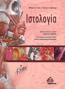
Ovalle, W.K. and P.C. Nahirney. Netter Ιστολογία. Greek translation: Philadelphia: Elsevier/Saunders 2011 (ISBN: 978-960-489-088-0), 500 pages.
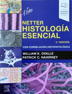
Ovalle, W.K. y P.C. Nahirney. Netter Histologia Esencial. Spanish translation: Barcelona, Spain: Elsevier, 2021 (ISBN: 978-849113-953-9), 551 pages. https://www.elsevier.com/books/netter-histologia-esencial/ovalle/978-84-9113-953-9
Web-related Materials
Ovalle, W.K. and P.C. Nahirney, Netter’s Histology Flash Cards for iPhone and iPad. Downloadable in US from https://itunes.apple.com/ca/app/netters-histology-flash-cards/id286993972?mt=8
Ovalle, W.K. and P.C. Nahirney. Online collection of zoomifiable electron micrographs from Netter’s Essential Histology. Elsevier/Saunders, Philadelphia 2008 and 2013 (StudentConsult.inkling.com).
Ovalle, W.K. and P.C. Nahirney. Online collection of 20 virtual slides . Part of web-related material for Netter’s Essential Histology. Elsevier/Saunders, Philadelphia 2008 (StudentConsult.inkling.com).
Ovalle, W.K. and P.C. Nahirney. Online Testbank of Multiple Choice Questions in Histology for Instructors. Part of web-related material for Netter’s Essential Histology. Elsevier/Saunders, Philadelphia 2008 (Evolve.elsevier.com).
Ovalle, W.K. and P.C. Nahirney. Video (Powerpoint) Overviews: 20 chapters. Part of web-related material for Netter’s Essential Histology, 2nd Edition. Elsevier/Saunders, Philadelphia 2013 (StudentConsult.com).URL: https://studentconsult.inkling.com/read/netters-essential-histology-ovalle-nahirney-2nd/videos/4fe0d8a615ab49708c63020ed7facc6c
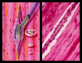
Schematic view (left) and H&E stained longitudinal section (right) of a muscle spindle.
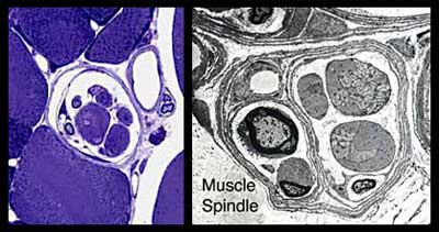
Transverse sections of muscle spindles. Toluidine Blue stained semi-thin section (left) and Transmission Electron Micrograph (right).
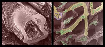
HRSEM view of a myelinated nerve fiber (left) and a mitochondrion in skeletal muscle (right).
