Ivan Robert Nabi
Professor
Cellular & Physiological Sciences
Faculty of Medicine
Other Titles
Director, LSI Imaging Facility
Contact
Email: ivan.robert.nabi@ubc.ca
Office: 604-822-7000
Lab: 604-822-7329
Education
PhD Cancer Metastasis (Weizmann Institute of Science)
BSc Biochemistry (McGill University)
Work in the Nabi lab focuses on the cell biology of cancer and the use of super-resolution microscopy to study cellular domains.
Research projects:
1) The role of non-caveolar scaffold domains and the galectin lattice in cancer cell migration and focal adhesion tension.
We study the role of plasma membrane domain effectors galectin-3 and caveolin-1 in focal adhesion dynamics and tension in metastatic cancer cells. We have developed network analysis of dSTORM super resolution microscopy to define the molecular architecture of caveolae and scaffolds and are now studying the structural changes in these membrane domains associated with tumor cell migration and mechanical stress. Our characterization of the proteome and transcriptome of tumor cell pseudopodia has identified multiple pseudopod-localized effectors whose role in pseudopod-specific actin dynamics and focal adhesion tension is being studied.
2) The role of the cancer-associated ubiquitin ligase Gp78 in the regulation of endoplasmic reticulum-mitochondria contacts and mitophagy.
We have defined the role of Gp78 (also known as autocrine motility factor receptor (AMFR)), a cancer-associated receptor and E3 ubiquitin ligase in endoplasmic reticulum (ER) associated degradation, in ER-mitochondria interaction and mitophagy. We have shown that Gp78 and its ligand AMF control rough ER-mitochondria contacts and are defining the underlying molecular mechanisms and applying STED super-resolution microscopy to define the organization of the ER and its interaction with mitochondria.
- Shapira T, Monreal IA, Dion SP, Jager M, Désilets A, Olmstead AD, Vandal T, Buchholz DW, Imbiakha B, Gao G, Chin A, Rees WD, Steiner T, Nabi IR, Marsault E, Sahler J, August A, Van de Walle G, Whittaker GR, Boudreault PL, Aguilar HC, Leduc R, Jean F. A novel highly potent inhibitor of TMPRSS2-like proteases blocks SARS-CoV-2 variants of concern and is broadly protective against infection and mortality in mice. Nature. 2022 In press.
- Timothy H. Wong*, Ismail M. Khater*, Bharat Joshi, Mona Shahsavari, Ghassan Hamarneh#, Ivan Robert Nabi#. Single molecule network analysis identifies structural changes to caveolae and scaffolds due to mutation of the caveolin-1 scaffolding domain. Scientific Reports. 2021 11:7810.
- Long RKM*, Moriarty KP*, Cardoen B, Gao G, Vogl AW, Jean F, Hamarneh G#, Nabi IR#. Super resolution microscopy and deep learning identify Zika virus reorganization of the endoplasmic reticulum. Sci Rep. 2020 Dec 1;10(1):20937. doi: 10.1038/s41598-020-77170-3. *,#equal contribution.
- Khater IM, Nabi IR*, Hamarneh G*. Super-resolution Single Molecule Localization Microscopy Cluster Analysis and Quantification Methods. Patterns. 2020 Jun 12;1(3):100038. doi: 10.1016/j.patter.2020.100038. eCollection 2020 Jun 12. *equal contribution.
- Cardoen B, Yedder HB, Sharma A, Chou KC, Nabi IR, Hamarneh G. ERGO: Efficient Recurrent Graph Optimized Emitter Density Estimation in Single Molecule Localization Microscopy. IEEE Trans Med Imaging. 2019 Dec 25.
- Caveolin-1 Y14 phosphorylation suppresses tumor growth while promoting invasion. Joshi B, Pawling J, Shankar J, Pacholczyk K, Kim Y, Tran W, Meng F, Rahman AMA, Foster LJ, Leong HS, Dennis JW, Nabi IR. Oncotarget. 2019 Nov 19;10(62):6668-6677.
- Gao G, Zhu C, Liu E, Nabi IR. Reticulon and CLIMP-63 regulate nanodomain organization of peripheral ER tubules. PLoS Biol. 2019 Aug 30;17(8):e3000355.
- Khater IM, Liu Q, Chou KC, Hamarneh G*, Nabi IR*. Super-resolution modularity analysis shows polyhedral caveolin-1 oligomers combine to form scaffolds and caveolae. Sci Rep. 2019 Jul 8;9(1):9888. *equal contribution.
- Khater IM, Meng F, Wong TH, Nabi IR*, Hamarneh G* (2018) Super resolution network analysis defines the molecular architecture of caveolae and caveolin-1 scaffolds. Scientific Reports 8, 9009. *equal contribution
- Fanrui Meng, Sandeep Saxena, Youtao Liu, Bharat Joshi, Timothy H Wong, Jay Shankar, Leonard J. Foster, Pascal Bernatchez, Ivan R. Nabi (2017). The phospho-caveolin-1 scaffolding domain dampens force fluctuations in focal adhesions and promotes cancer cell migration. Mol Biol Cell. 2017 28, 2190-2201
Further publications can be found here.
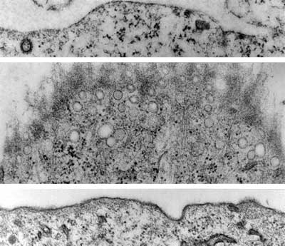
Plasma membrane domain organization regulates EGFR signaling in tumor cells. Cover image, Journal of Cell Biology 179, 2007.